Subperiosteal implants
1. Diagnosis and Digital Mapping
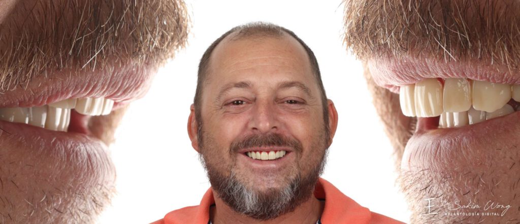
The process begins with high-resolution facial scanning, intraoral digital capture, and cone-beam tomography. This combination allows us to visualize both external anatomy and internal skeletal structures, accurately assessing bone volume, density, and maxillary morphology before any intervention begins.
2. Custom Implant Engineering
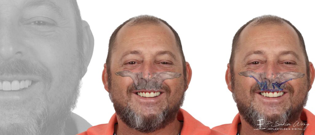
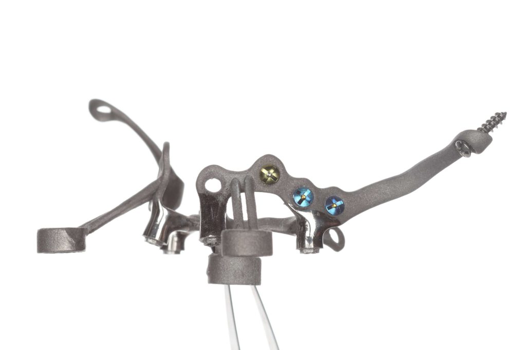
Using the combined scan and tomography data, we engineer a patient-specific subperiosteal framework designed to match the surface anatomy of the maxilla with controlled fixation points and optimized load distribution.

3. Prosthetic Planning and Simulation
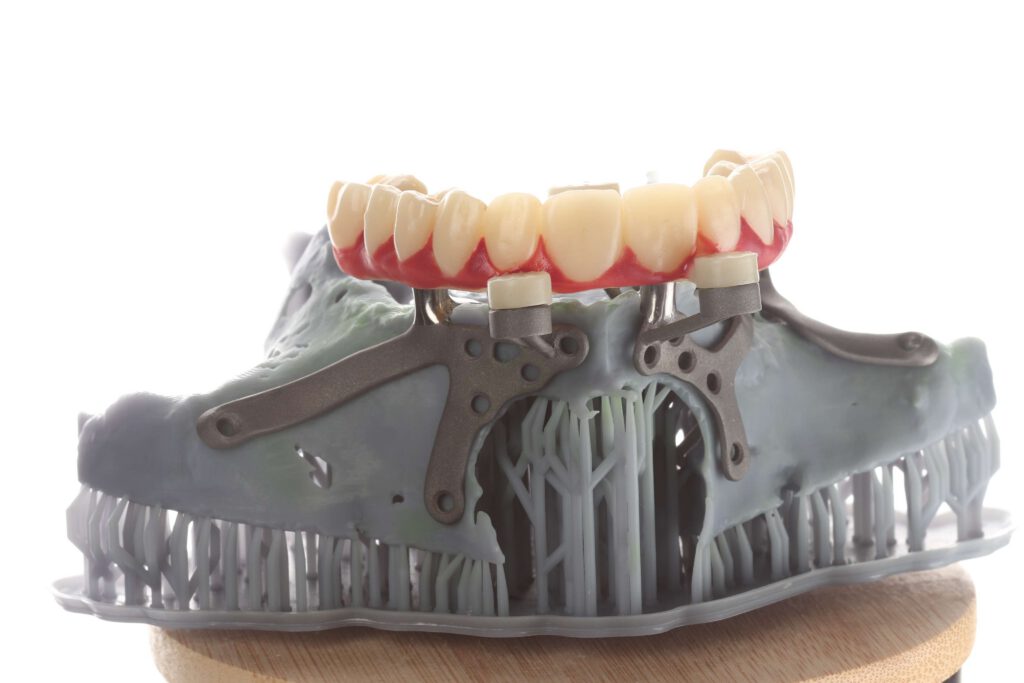
The implant is integrated into a 3D bone model to validate fit, biomechanics, and prosthetic positioning. Temporary prosthetic components are designed in parallel to maintain functional continuity from surgery to final restoration.
4. Precision Manufacturing
Medical-grade titanium is manufactured for the implant framework, while zirconia is selected for strength, biocompatibility, and aesthetics in prosthetic components. Every interface is refined for long-term biological integration and mechanical performance.
5. Surgical Execution and Final Result
The implant is placed with high precision, followed by the installation of a provisional prosthesis to restore function early. Once integration is achieved, the final zirconia prosthesis is delivered—transforming complex anatomy into stable function and natural aesthetics.